Peripheral inflammation may drive Parkinson's, study finds
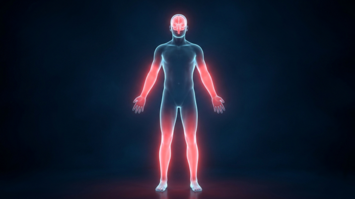
New research suggests peripheral inflammation may drive Parkinson's disease: aging or LRRK2 mutations spark low-grade immune activity outside the brain, and DNA-carrying extracellular vesicles ferry those signals across a leaky blood-brain barrier to activate microglia and erode motor function. Scientists report the damage starts in the body before it reaches the brain.
Key Takeaways
- A Cell Reports study links peripheral inflammaging to Parkinson's via STING-dependent interferon signaling.
- LRRK2 G2019S mutations and normal aging both boost DNA-filled extracellular vesicles that spread inflammation.
- Peripheral IFN-I rose by 3 months in mice, but brain inflammation and motor decline appeared at 12 months.
- Deleting STING normalized immune responses and prevented dopaminergic neuron loss in Lrrk2 mutant mice.
- Human Parkinson's samples showed elevated DNA-containing vesicles and STING-driven interferon activity.
What Did the New Parkinson's Study Find?
An international team published findings in Cell Reports suggesting that STING-dependent peripheral inflammaging can fuel neurodegeneration through extracellular vesicles. The researchers tested plasma and cerebrospinal fluid from young donors, aged healthy donors, and Parkinson's patients.
Parkinson's patients showed elevated systemic type I interferon activity, but not NF-κB signaling. Blood monocytes from those patients carried higher IFNB1 transcripts, and an LRRK2 inhibitor brought those levels back toward healthy ranges. Several human experiments used small sample sizes of just three to four participants.
How Do Extracellular Vesicles Spread Inflammation to the Brain?
The study centers on the LRRK2 G2019S mutation, the most common genetic cause of Parkinson's. This gain-of-function change disrupts endolysosomal recycling, letting self-DNA from damaged nuclei or mitochondria spill into the cytosol and get packaged into circulating extracellular vesicles.
Those vesicles can activate the cGAS-STING pathway in distant cells, including immune cells and eventually brain microglia. In G2019S knock-in mice, plasma vesicle buildup appeared early, while cerebrospinal fluid levels rose much later, matching a peripheral-first timeline.
What Role Does the STING Pathway Play?
The cGAS-STING pathway normally detects foreign DNA, but aging and senescence can trigger the same alarm with a cell's own genetic material. That produces a chronic type I interferon response often called inflammaging, a term coined by Claudio Franceschi to describe age-linked systemic inflammation.
In Lrrk2 mutant mice, deleting STING fully reversed elevated interferon signaling in both splenocytes and microglia. STING loss also cut microglial inflammation markers, protected against age-related motor decline, and blocked the 51% dopaminergic neuron loss seen in mutants versus about 30% in wild-type mice.
Why Does This Matter for Longevity Research?
The findings reinforce a growing view that brain aging is not sealed off from the rest of the body. A leaky blood-brain barrier may be the gateway that lets peripheral immune chatter reshape microglial behavior over time. For readers tracking broader anti-aging science, see our Longevity & Biohacking coverage.
Related work on clearing senescent cells and supporting repair pathways continues to advance in parallel. A recent mouse study pairing senolytics with stem cells hints at how dampening SASP inflammation might complement strategies targeting systemic immune drivers like STING.
Prior Nature research on cGAS-STING also tied this pathway to age-related inflammation and neurodegeneration. Researchers caution that human sample sizes were limited and more validation is needed before clinical translation.