Exosomes from stem cells reverse liver aging signs in mice
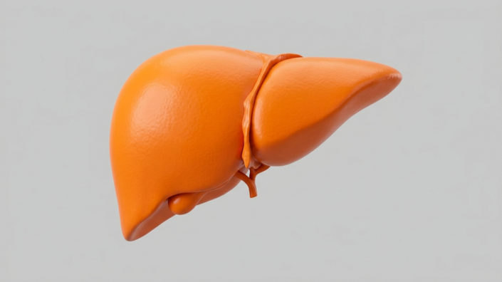
Exosomes from stem cells taken from human umbilical cord tissue improved liver function and lipid markers in aged male mice, Lifespan.io reports. The tiny vesicles raised autophagy, lowered senescence, and shifted key blood and liver biomarkers toward youthful levels via the protein THBS1.
Key Takeaways
- Human umbilical cord mesenchymal stem cell exosomes (HucMDEs) reached mouse livers after tail-vein injection.
- Treated old mice showed better ALT, AST, triglycerides, cholesterol, glucose, and body-weight trends versus untreated aged controls.
- Effects tracked with more autophagy, less senescence, and recovery of fat-handling proteins such as PPARα.
- The cargo protein THBS1 was required; without it, the exosomes lost their anti-aging liver benefits.
- Findings remain limited to male mice and are not yet a clinical therapy for NAFLD.
What did the stem cell exosome study actually find?
According to Lifespan.io, researchers used exosomes from human umbilical cord mesenchymal stem cells (HucMSCs) to counter age-linked metabolic stress in the liver. After confirming the parent cells could become bone and fat cells, they harvested exosomes, labeled them, and injected them into mouse tail veins. Marker tracking showed the vesicles were taken up in liver tissue.
Animals included young (8-week-old) males, untreated 18-month-old males, and old males given HucMDEs. Untreated old mice were much heavier and had sharply higher blood glucose. Treated old mice were only slightly heavier than young animals, and their glucose looked nearly like the young group.
Four fatty-liver markers moved in the same direction. ALT and total cholesterol in treated animals returned toward young levels. AST and triglycerides also fell significantly, though not fully to youthful baselines. Lipid deposits in the liver dropped, SREBP1 fell, and PPARα partly recovered. Researchers wrote that aging drove liver dysfunction and dyslipidemia, and that HucMDE dosing could improve function and normalize serum lipids in aging mice.
How do exosomes from stem cells protect liver cells?
The work centered on autophagy, the cleanup process that clears faulty organelles and fatty droplets in hepatic cells. Weak autophagy is linked to non-alcoholic fatty liver disease (NAFLD), and boosting it has previously reduced liver injury in mice. HucMDEs raised autophagy markers such as LC3 in cells and animals, with RNA-silencing work confirming higher autophagic flux.
Senescence signals also eased. Liver levels of p16 and p21 in treated old mice resembled those in young mice. In cultured liver cells stressed with palmitic acid, HucMDEs—but not lung-fibroblast exosomes—cut the senescence marker SA-β-Gal and improved fat-related readouts.
Those benefits depended on THBS1, a protein abundant in HucMDEs but not in fibroblast exosomes. Knocking down THBS1 before packing the vesicles wiped out effects on autophagy and senescence. Blocking PPARα in target cells likewise blunted useful HucMDEs, tying the pathway together.
Why does this matter for longevity and biohacking?
Readers following longevity and biohacking research care because NAFLD still lacks a specific approved drug, and age-related liver fat plus cellular senescence are major metabolic risks. MSC-derived exosomes have also been explored against sarcopenia and other liver conditions, so this study adds a clearer mechanism—THBS1 acting through PPARα—rather than a vague “stem cell” claim.
Caveats are essential. The experiments used only male mice. Autophagy recovery was incomplete versus young animals. Human trials are not described in the source coverage. Parallel geroscience work, such as enzyme strategies against age-related protein damage, shows how many repair angles are still preclinical. For now, exosomes from stem cells look like a promising experimental lever against aging liver dysfunction—not a consumer therapy.