ALS microglia eat living neurons, mistaking them for dead
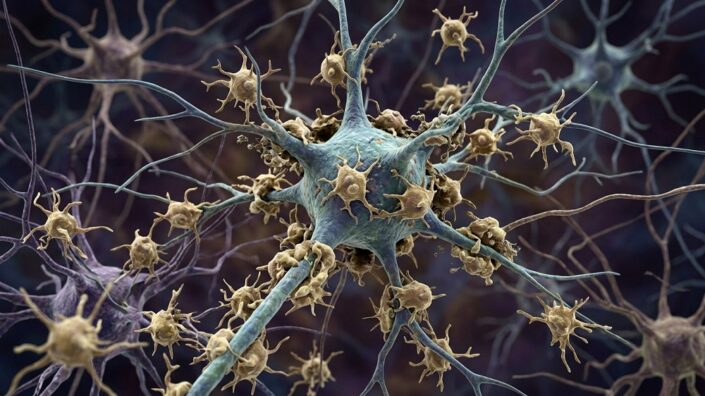
ALS microglia eat living motor neurons after mistaking stress signals for death cues. A new Nature Communications study found elevated TAM receptors AXL and MER drive this misfire, and blocking them in mice preserved neurons and delayed the disease endpoint.
Key Takeaways
- Stressed motor neurons in ALS can display phosphatidylserine, an “eat-me” signal normally linked to dying cells.
- Brain immune cells called microglia use TAM receptors AXL and MER to recognize that signal and engulf living neurons.
- Human ALS spinal cord tissue showed roughly threefold higher MER and about 16-fold higher AXL than controls.
- In SOD1 ALS mice, silencing Axl and Mer preserved about three times as many motor neurons and cut neuronal debris in microglial lysosomes roughly tenfold.
- Similar TAM activation has been seen in Alzheimer’s and Parkinson’s models, hinting at a broader neurodegeneration pathway.
Amyotrophic lateral sclerosis is an age-related disease marked by progressive motor-neuron loss. Much research has focused on defects inside those neurons. Growing evidence shows neighboring cells matter too—especially microglia, the brain and spinal cord’s resident immune cells. For more coverage of aging biology, see our Longevity & Biohacking hub.
Why do ALS microglia eat living neurons?
Microglia normally clear damaged or dead cells. They do that partly through TAM receptors AXL and MER, which recognize phosphatidylserine (PtdSer) flipped to a cell’s outer membrane during apoptosis.
A team led by Salk Institute’s Greg Lemke asked whether that cleanup system goes wrong in ALS. “Cells that are dying throw an ‘eat me’ sign out on their surface, and the TAM system recognizes that sign,” Lemke said. The group wondered whether microglia corrupt TAM signaling to kill living neurons.
In postmortem lumbar spinal cord from six people with sporadic ALS versus three age-matched controls, microglial MER was about threefold higher and AXL about 16-fold higher. SOD1G93A mice showed rising Axl and Mer in spinal microglia as disease advanced. Motor-neuron GAS6, a bridge between PtdSer and TAM receptors, was also higher in ALS tissue and mice. Late-stage SOD1 mice had many PtdSer-positive motor neurons, yet most lacked cleaved caspase-3, a marker of ongoing apoptosis—suggesting the “eat-me” display came from still-living cells.
What happened when researchers blocked the eat-me pathway?
Crossing SOD1 mice with animals lacking both Axl and Mer produced a striking pattern: earliest illness appeared about 30 days sooner, but the terminal clinical endpoint arrived roughly three weeks later. Those mice kept about three times as many motor neurons, with better axon–muscle connections and preserved muscle size.
Because whole-body TAM loss can cause autoimmunity and other problems earlier, the team also deleted the receptors mostly in adult microglia after disease onset. That conditional silencing still extended survival: by day 160, none of the treated animals had reached the clinical endpoint, versus about 60% of controls. Microglial lysosomes in ordinary SOD1 mice held abundant neuronal material; Axl/Mer deletion reduced it by roughly tenfold.
“The bottom line is, microglia are using the TAM system to eat cells that aren’t dead,” Lemke said, noting potential for TAM-targeted approaches that are simpler than engineering whole-cell immunotherapies. First author Youtong Huang reported that losing the TAM proteins meant preserving muscle control in the model.
Could this matter beyond ALS?
The findings are detailed in Nature Communications and summarized by Lifespan.io. Very similar TAM activation has been observed in Alzheimer’s and Parkinson’s disease models. Authors therefore speculate that PtdSer-dependent killing of stressed-but-living neurons by microglia may be a general neurodegeneration mechanism—not ALS alone. Human trials are still needed before any clinical claim.